The crustcerebral is the newest (evolutionarily) and largest part of the brain. This is where perception, imagination, thought, judgment, and decision occur. It is the largest region of the mammalian brain and plays a key role in memory, attention, perception, cognition, consciousness, thought, language, and consciousness.
In the following Psychology-Online article, we will explain in detail what is the cerebral cortex, what are its layers, parts and functions.
Index
- What is the cortex or cerebral cortex - definition and function
- Layers of the cerebral cortex
- The cerebral hemispheres
- Left and right hemisphere: experiments on epilepsy
- Language and the cerebral cortex
- Photos about the cerebral cortex
What is the cortex or cerebral cortex - definition and function.
The cerebral cortex is primarily a thin gray matter layer - typically 6 neurons thick, in fact - above a vast collection of white matter pathways. The thin layer is strongly convoluted, so if you spread it, it would occupy about 2500 cm2. This layer includes about 10 billion neurons, with about 50 trillion synapses. The convolutions have "ridges," which are called gyri, and "valleys," which are called furrows.
Parts and functions of the cerebral cortex
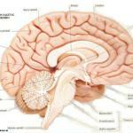
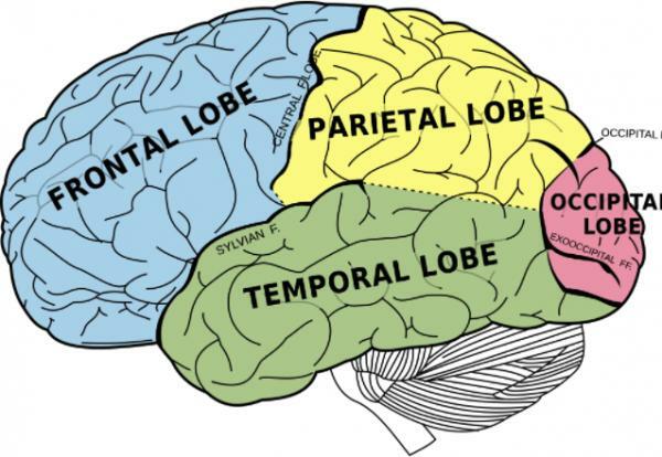
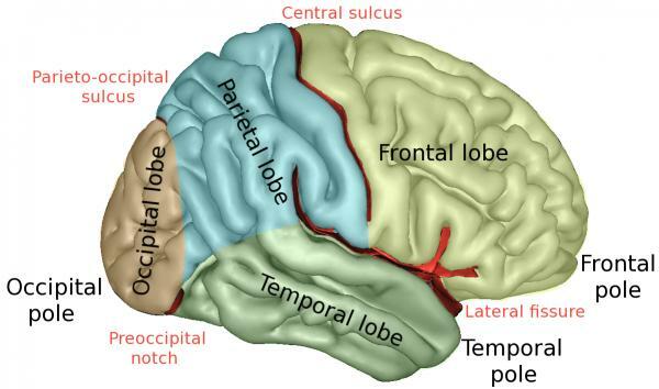
Some furrows are quite pronounced and long and are used as agreed boundaries between the four areas of the brain called lobes. The furthest front part is called the frontal lobe. This seems to be especially important: this lobe is responsible for the movements volunteering and planning and is thought to be the most important lobe for personality and intelligence.
At the back of the frontal lobe, along the sulcus that separates it from the parietal lobe, there is an area called motor cortex. In studies with patients undergoing brain surgery, stimulation of areas of the motor cortex with small electrical shocks caused movements. It has been possible for researchers to make a fairly accurate map of our motor cortex. The lower parts of the motor cortex, near the temples, control the muscles of the mouth and face. The parts of the motor cortex near the top of the head control the legs and feet.
Under the frontal lobes is the parietal lobe (which is Latin for "wall"). This includes an area called somatosensory cortex, just below the sulcus that separates this lobe from the frontal lobe. Again, the doctors stimulated the points in this area, finding that their patients described sensations as if they were being touched in various parts of their body. As with the motor cortex, the somatosensory cortex can be mapped, with the mouth and face near the temples and the legs and feet on top of the head.
Next to the head is the temporal lobe (it is the Latin term for "temples"). The special area of the temporal lobe is the auditory cortex. As the name suggests, this area is intimately connected with the ears and specialized in the ear. It is located near the connections of the temporal lobe with the parietal and frontal lobes. At the back of the head is the occipital lobe. At the rear of the occipital lobe is visual cortex, which receives information from the eyes and specializes, of course, in vision. The areas of the lobes that are not specialized are called association cortex. In addition to connecting the sensory and motor cortices, this is also thought to be the place where our thought processes occur and many of our memories are ultimately stored.
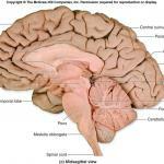
Below we offer you an image so that you know the main parts of the cerebral cortex
Layers of the cerebral cortex.
There are 6 layers of the cerebral cortexNext, we are going to explain each one of them, ordered from the surface to the inside of it:
- Molecular layer: Also called the plexiform layer, it is the most superficial layer of the cerebral cortex. It is a synaptic layer composed of a dense network of nerve fibers, which derive pyramidal and spindle cell dendrites, the axons of Martinotti cells and starred. Being the outermost layer, in it, many synapses are established between neurons.
- Outer granular layer: It is located below the molecular layer and, in it, there are many small stellate and pyramidal cells. The dendrites of cells and axons infiltrate deeper layers, so this layer is interconnected with the different parts of the cortex.
- Outer pyramidal layer: It is made up of pyramidal cells and has an irregular shape with a size that increases from the surface to the deepest part. Pyramidal cells direct their axons to other parts of the cortex in the form of projection, association, or commissural fibers.
- Inner granular layer: It is made up of the stellate cells, which are arranged in a compact way. It has the outer Baillarger band, which are fibers arranged horizontally.
- Inner pyramidal layer: Also called ganglionic layer, it has pyramidal cells of medium and large size. It has a large number of fibers arranged horizontally, which make up the so-called internal Baillarger band.
- Multiform or polymorphic layer: It is made up of spindle cells and also has modified pyramidal cells, which have a triangular or ovoid body. The nerve fibers of this layer of the cerebral cortex enter the underlying white matter connecting with the intermediate regions. The spindle cells derive information into the cortex, thalamus, and striated nuclei.
The cerebral hemispheres.
If you look at the brain from above, it becomes immediately obvious that there is a split in two from front to back. There are, in fact, two hemispheres, as if we have two brains in our heads instead of just one. Of course, those two halves are intimately linked by an arc of white matter called hard body. In various ways, researchers have found that the two parties have some specialization.
Functions and differences of the cerebral hemispheres
The left hemisphere is related to the right side of the body (usually), and the right hemisphere it is related to the left part of the body. In addition, it is the left hemisphere that normally has language, and it seems to be the main responsible for similar systems such as mathematics and logic. The right hemisphere has more to do with things like spatial orientation, face recognition, and body image. It also seems that he governs our ability to appreciate art and music. Some of the most interesting work that has been done related to the two hemispheres was carried out Roger sperry. He worked with people who had had a serious enough operation to control his epilepsy.
The right side of each retina (which sees things to the left of the fixation point) goes to the left hemisphere. What this means is that if you have someone staring at one fixation point and you briefly show them something to the left, it is the right hemisphere that receives the information. If you show them something to the right, it is the left hemisphere that receives the information. Sperry would project things onto a screen and ask patients to either say what they had seen or to take what they had seen with one hand or the other from a box full of things. Thus, if the he showed a ball on the left side of the screen and a pencil on the right, the person could say "pencil" (using the language centers of the left hemisphere) but catch a ball from the box with his left hand (using the left hemisphere right).
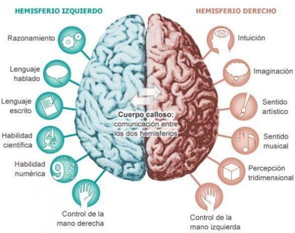
Image: areaciencias.com
Left and right hemisphere: experiments on epilepsy.
It seems that, in some cases, severe epilepsy can be almost eliminated sectioning the corpus callosum. In a sense, these people actually had two brains (or cortices, to be more exact).
For example, Sperry[1] he found that if he put something on the right hand of one of these people after his operation, they could tell what it was. But if he put it in his left hand, they couldn't do it. This is easy to understand: The sensation of an object in the right hand goes to the left hemisphere and, since this is the language zone, the person could tell what it was. The sensation of a thing in the left hand, however, was going to the right hemisphere, which cannot speak much. The eyes are connected to the hemispheres in a bit of a complicated way.
To this day, it is often questioned whether the functions are really so separated by hemispheres or rather they are spread throughout the cerebral cortex.
Language and the cerebral cortex.
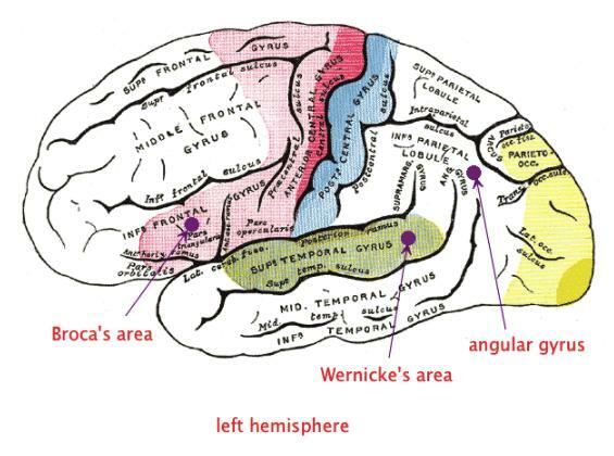
Therefore, language is predominantly a function of the left hemisphere. Actually, the right hemisphere has a bit of language too: it has a good understanding of insults and curse words. Also, if you have brain damage in the left hemisphere early enough in childhood, the right hemisphere takes over language function. And it seems that there are some people who have the language on the right side or even on both sides. It is interesting to consider that monkeys and gorillas seem to be sensitive to calls of their own species in the left hemisphere: they turn their right ears towards the sound. Even some songbirds, like canaries, have hemispheric specialization. One of the earliest discovered things about the brain was the language centers.
One of them is called the Broca's area, on behalf of the doctor who discovered it first. It is located in the lower part of the left frontal lobe. A patient who has had damage in that area loses the ability to speak, which is called expression aphasia. Another area is the area of Wernicke, which is close to Broca's area but in the temporal lobe, right next to the auditory cortex. This is where we understand the meaning of language, and damage in this area would lead to a reception aphasia, which means that you would not be able to understand what is being said to you.
Occasionally, someone has a damage to the connections between the Wernicke and Broca areas. This leads to a conduction aphasia. Some people with this problem can understand language quite well, and they can produce it just as well. But they can't repeat something they just heard. Another important area is the turn angular, just above and below Wernicke's area. It serves as a connection between the language centers and the visual cortex. If this area is damaged, the person will suffer from alexia (inability to read) and agrafia (inability to write).
Photos about the cerebral cortex.
If you want to know more about the functions of the cerebral cortex and nervous system anatomy we offer you the following article: parts of the brain and their functions.
Next, we also show you a battery of photographs and images about the cerebral cortex so that you can observe and study it.
Image source: snowbrains and pinterest
This article is merely informative, in Psychology-Online we do not have the power to make a diagnosis or recommend a treatment. We invite you to go to a psychologist to treat your particular case.
If you want to read more articles similar to The cerebral cortex: functions and parts, we recommend that you enter our category of Cognitive psychology.
References
- Myers, R. E., & Sperry, R. W. (1958). Interhemispheric communication through the corpus callosum: mnemonic carry-over between the hemispheres. AMA Archives of Neurology & Psychiatry, 80(3), 298-303.
Bibliography
- Restrepo, F. J. L. (2008). Executive functions: clinical aspects. Journal of Neuropsychology, Neuropsychiatry and Neurosciences, 8(1), 59-76.
Photos of The cerebral cortex: functions and parts