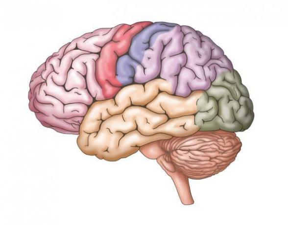
Image: Feel the Brain
One day you start to feel bad and suddenly it is difficult for you to perform some movements that you used to do without any problem. It may happen that you stop feeling pain, discomfort or sensitivity in a particular area of the body such as the fingers, the face, the arms, among others. Your daily activities are directly affected by these inconveniences. Maybe it happened to you at some point in your life or it happened to someone you know. Our body gives us signals that we must listen carefully to deal with them. There are some diseases such as epilepsy that influence the performance of the brain. However, we currently have ways of studying our brain to see how it works. Do you want to know more about this? In this Psychology-Online article, we will provide you with information about what is the sensory and motor Penfield homunculus.
Index
- Penfield homunculus story
- Function of the Penfield homunculus
- The two cerebral homunculi
History of the Penfield homunculus.
The Penfield homunculus has its origin in the studies and developments carried out by Dr. Wilden Penfield about graphic representations of the brain. This doctor was looking for ways to cure some neurological diseases such as epilepsy, among others.
During the year 1928, Dr. Penfield worked with Otfrif Foerster in the creation of a method to study different brain areas. There were certain patients who presented brain injuries caused by various diseases and / or accidents, in such a way that electrodes were placed in different sectors of the head. Then, small electric shocks were sent to them in order to know if some areas of the cerebral cortex were responding to these stimuli. This resulted in the doctors knowing whether or not there were affected areas that could be removed.
Why is the Penfield homunculus study important? These studies allowed Penfield to develop a graphic representation of the brain which shows that there is a connection between different parts of the human body, the body's sensitivity and the neurons that carry information that is internally decoded.
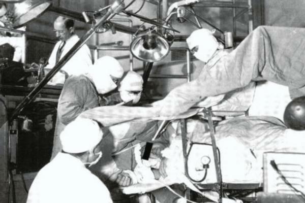
Image: Somatosphere
Function of the Penfield homunculus.
What does the homunculus represent? The Penfield homunculus gives us the possibility of accessing a body map in which both the motor area and the sensitive area of the body are represented human. This chart is used by physicians specialized in neuropsychiatry for the diagnosis and treatment of certain diseases with a neurological basis that can cause damage to different specific areas of the cortex cerebral. For this reason, the approach to this type of problem should be in charge of a health professional since the knowledge and experience that he possesses enable him to indicate an adequate treatment according to the characteristics of the patient. It is important that we know that there are variables that influence the clinical diagnosis such as age, sex, family history and pre-existing diseases of the patient, among others.
Next, we will see what is and where is Penfield's motor and sensitive homunculus.
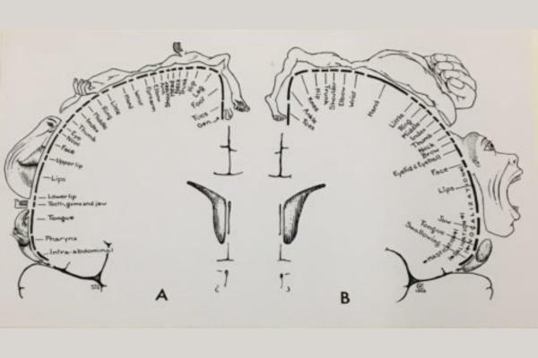
Image: Somatosphere
The two cerebral homunculi.
exist two types of cerebral homunculi that we need to distinguish since they have different functions and characteristics from each other. Next, we will see the location of the sensitive and motor Penfield homunculus and describe the most important qualities of each of them:
What is the motor homunculus and where is it found?
The motor homunculus corresponds to the regulation and control of body movements. It is located in the center of the frontal cortex, which includes the frontal lobe of the brain. The motor homunculus simultaneously receives and sends sensory information that is used for planning and executing movements. The graphic representation of the motor homunculus includes various areas of the human body such as face, fingers, and arms, among others. In addition, the functions of each of these parts of the body such as swallowing and blinking also appear. If we look at the graph of the Penfield motor homunculus, we can see that there are some areas larger than others, beyond the actual size of each sector. This is because the larger the area represented, the stronger the neuronal connections and the more space it occupies within the cerebral cortex.
What is the sensory homunculus and where is it found?
The sensitive homunculus is the brain area responsible for graphically represent the body's sensitivity to touch, pressure, and pain that a person can experience. Its location consists of the parietal lobe in the area that joins the frontal lobe. This area sends and receives information largely through the thalamus, which is responsible for the integration of different sensations that are perceived. In this way, it is possible to obtain a variety of stimuli and decode them in the body thanks to the neural connections found in this area of the brain.
In this article, you will find more information about Parts of the brain and their functions.

Image: Cajal Neurosciences
This article is merely informative, in Psychology-Online we do not have the power to make a diagnosis or recommend a treatment. We invite you to go to a psychologist to treat your particular case.
If you want to read more articles similar to What is the sensory and motor Penfield homunculus, we recommend that you enter our category of Neuropsychology.
Bibliography
- Gordillo León, Mestas Hernández, L. (May 29, 2020). Lthe traces of our evolution in the brain: Penfield's homunculus. Recovered from: https://www.neuromexico.org/2020/05/29/las-huellas-de-nuestra-evolucion-en-el-cerebro-el-homunculo-de-penfield/
- Pesudo, J. V., González-Darder, J.M. (2004). Electrical stimulation of the motor cortex for the treatment of central pain and peripheral pain due to deafferentation. Spanish Society of Pain Magazine, 11 (7), 370-379.
- Sallés, L., Gironés, X., Lafuente, J. V. (2013). Motor organization of the cerebral cortex and the role of the mirror neuron system. Journal of Clinical Medicine, 144 (1), 30-34.